The winners of the 2016 Nikon Small World Photomicrography Comp. have been announced.
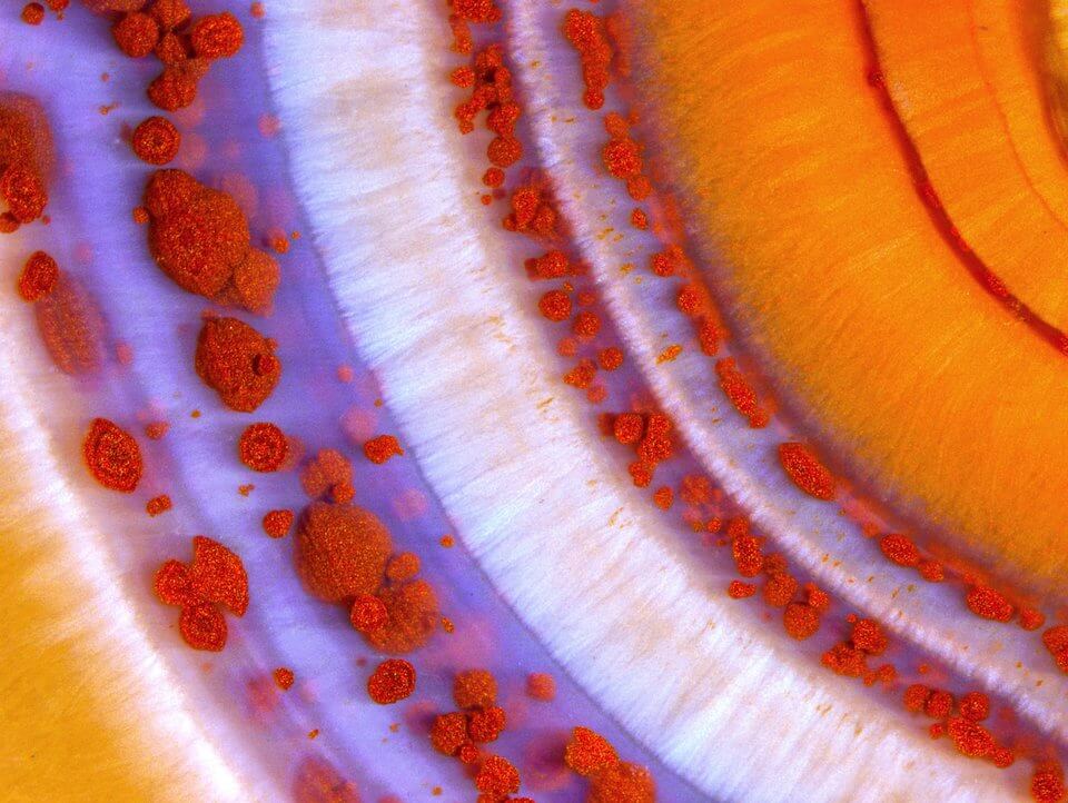
The competition showcases “the beauty and complexity of life as seen through the light microscope,” according to Nikon’s website. The pic above was shot by Douglas L. Moore of the University of Wisconsin, Stevens Point. It shows a polished slab of Teepee Canyon agate shown at 90x magnification.
In our role as brand designers we are constantly searching for the stunningly unique, and these pics provide that in spades. 96 stunning pics were honored in this year’s contest, these are twelve of my favorites.
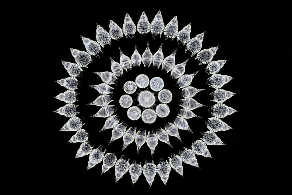
Stefano Barone, Palazzo Pignano, Italy. Sixty-five fossil Radiolarians (zooplankton) carefully arranged by hand in Victorian style. 100x magnification.
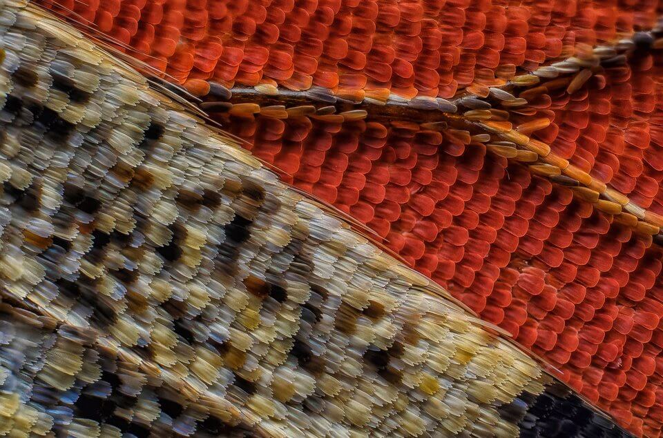
Francis Sneyers, Brecht, Belgium. Scales of a butterfly wing underside (Vanessa atalanta) shown at 10x magnification.

Marek Mis, Suwalki, Podlaskie, Poland. Air bubbles formed from melted ascorbic acid crystals shown at 50x magnification.
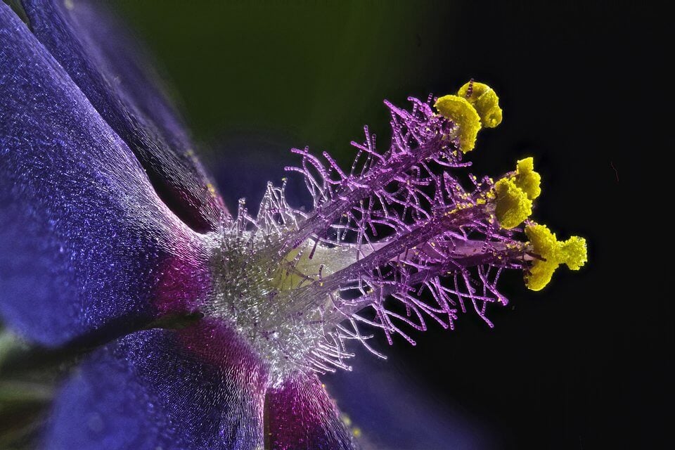
Samuel Silberman, Yehud-Monosson, Israel. Wildflower stamens shown at 40x magnification.

Jochen Schroeder, Chiang Mai, Thailand. Butterfly proboscis shown at 6.3x magnification.

Dr. Igor Siwanowicz of the Howard Hughes Medical Institute, Janelia Research Campus, Ashburn, Virginia. Front foot (tarsus) of a male diving beetle shown at 100x magnification.
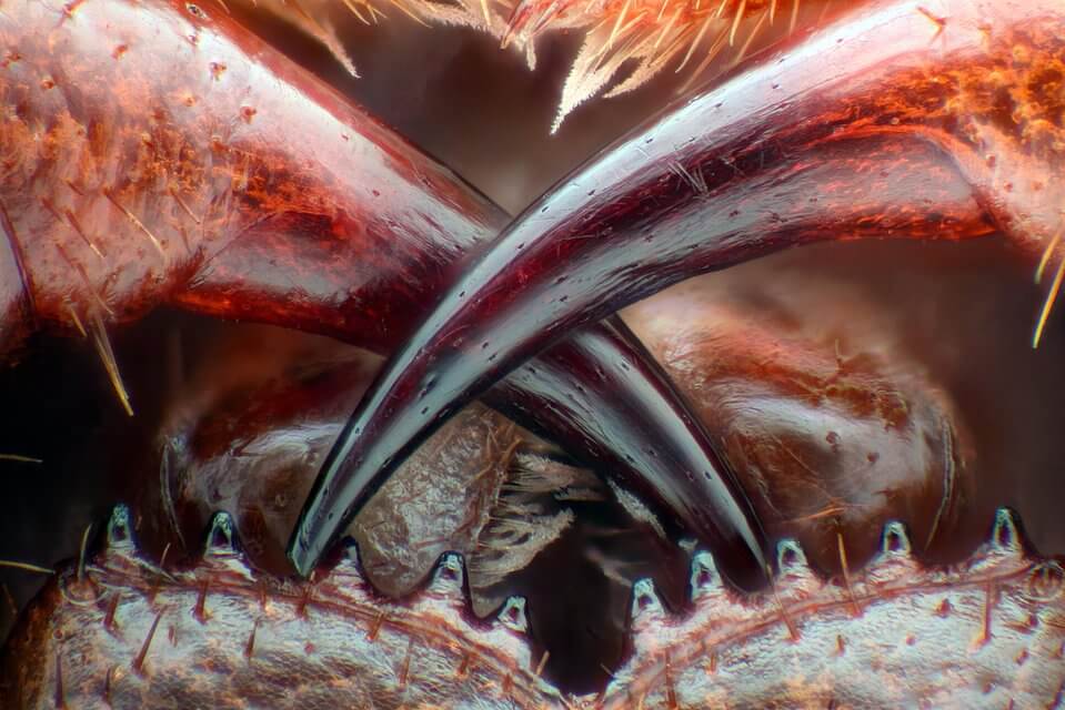
Walter Piorkowski, South Beloit, Illinois. Poison fangs of a centipede (Lithobius erythrocephalus) shown at 16x magnification.
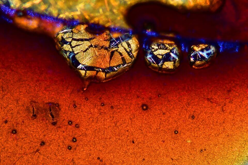
Vin Kitayama and Sanae Kitayama, Vinsanchi Art Museum, Azumino, Nagano, Japan. Espresso coffee crystals.
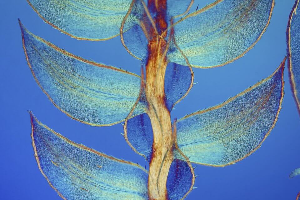
Dr. David Maitland of Feltwell, England. Leaves of Selaginella (lesser club moss) shown at 40x magnification.
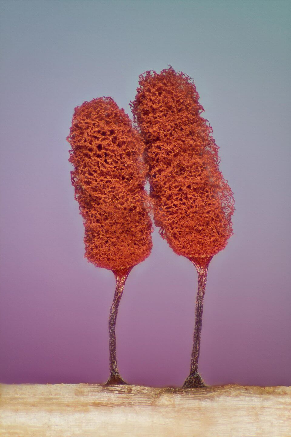
Jose Almodovar of the University of Puerto Rico, Mayaguez. Slime mold (Mixomicete) shown at 5x magnification.

Rogelio Moreno Gill, Panama, Panama. Paramecium-like Frontonia organism (showing ingested food, cilia, mouth and trichocysts) shown at 200x magnification.